

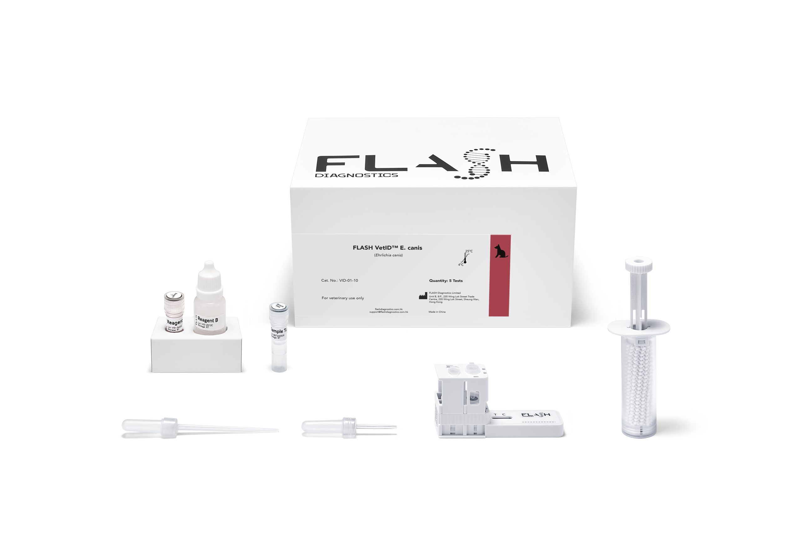
FLASH VetIDTM E. canis
Fast test for the qualitative detection of DNA of Ehrlichia canis in whole blood of the dog.
- Sensitivity 100.00% & specificity 100.00% (n=153)
- Detects DNA directly – the sensitivity of morulae microscopy can drop to single-digit percentages
40
minutes
Total assay time
50 μL
EDTA whole blood
Sample
Room
temperature
Storage
12
months
Shelf life
Clinical suspicion:
- Fever, lethargy, anorexia, lymphadenomegaly or splenomegaly, especially with tick exposure
- Bleeding tendencies: petechiae, ecchymoses or epistaxis
- Ocular signs: uveitis, chorioretinitis, or retinal detachment
- Chronic, unexplained weight loss, pancytopenia, or persistent weakness (chronic-phase presentation)
- Dogs from or residing in warm, high tick-exposure environments
Screening / prevention programs:
- Blood donor screening
- Dogs recently imported from or residing in endemic regions
- Kennel or shelter screening where tick exposure is uncontrolled
- Monitoring dogs with prior confirmed infection or incomplete treatment response
Pathogen and transmission
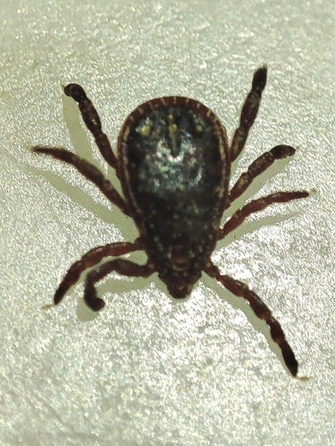
Ehrlichia canis is an intracellular bacterium infecting monocytes and the principal cause of canine monocytic ehrlichiosis. Transmission is mainly via the brown dog tick complex, and the disease is more common in tropical and subtropical regions and in kennel or household exposure settings. Infection can progress through acute, subclinical and chronic phases, though the boundaries between them are often indistinct in naturally infected dogs.
Species-Specific Manifestations
Acute-phase signs include fever, depression, reduced appetite, lymphadenomegaly and splenomegaly, with petechiae or epistaxis suggestive of thrombocytopenia. Ocular and renal involvement can occur even in subclinically infected dogs. The severe chronic phase is fundamentally a bone marrow suppression syndrome – pancytopenia, haemorrhage, and secondary infection are the principal drivers of death, and case fatality figures vary enormously between referral-hospital case series and general practice populations.
The Diagnostic Value Proposition
Blood smear detection of morulae is poor – present in lower than 10% of clinical-case smears. PCR is most reliable early in the acute phase, before antimicrobial treatment starts, since even limited prior doxycycline exposure can suppress detectable bacteraemia. Serology has its own gaps: antibodies typically take 12–14 days to become detectable, and a single positive titre cannot distinguish current from past infection. Direct DNA detection avoids both limitations, giving a same-visit, species-specific answer rather than waiting on a paired serology titre.

Overall workflow
1. Prepare the sample in 10 minutes with the SampleDirect cartridge
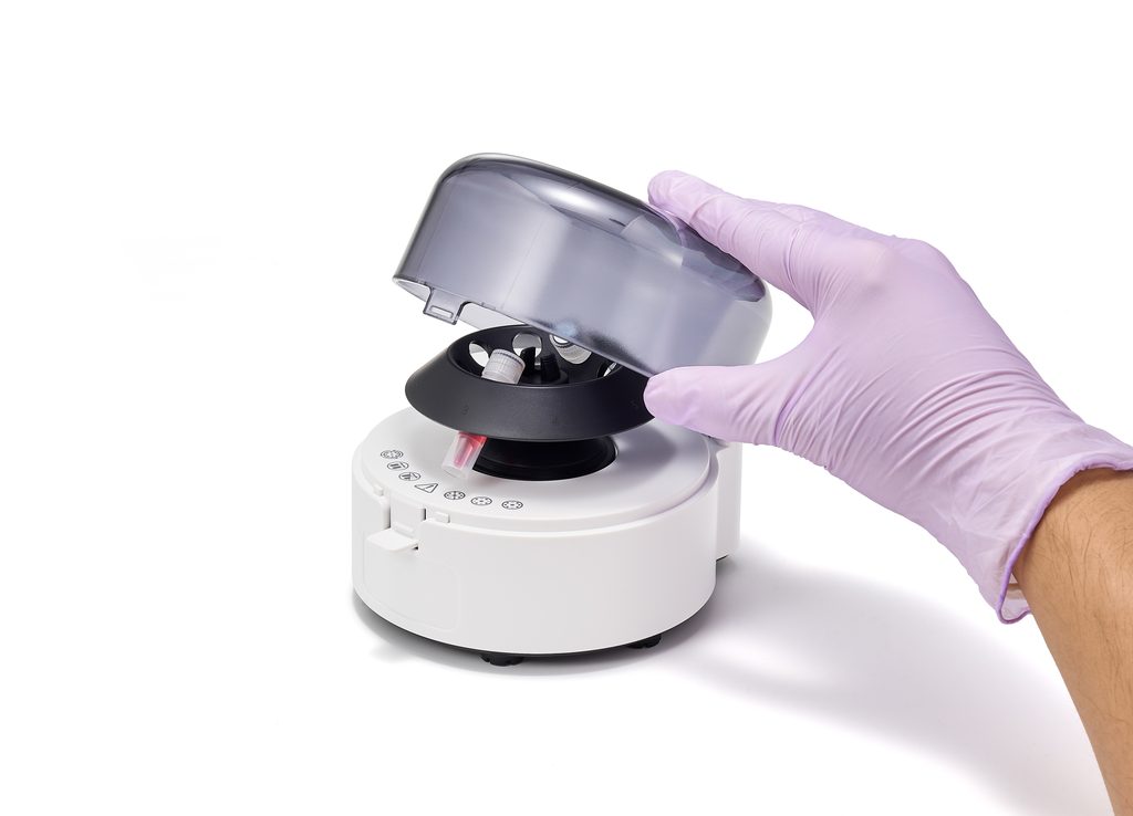
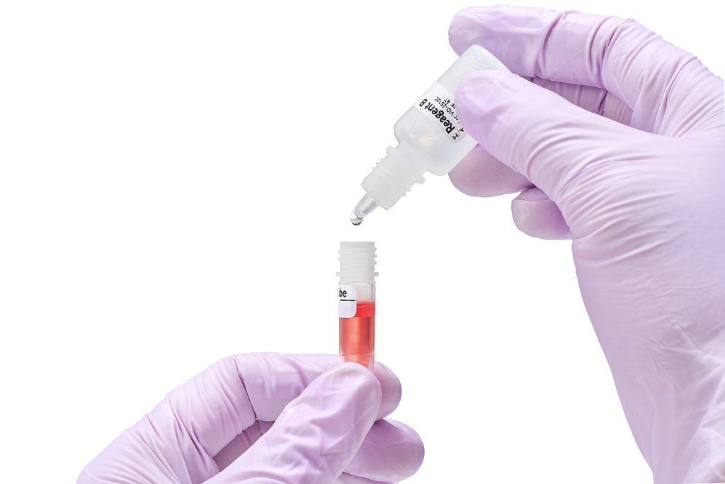
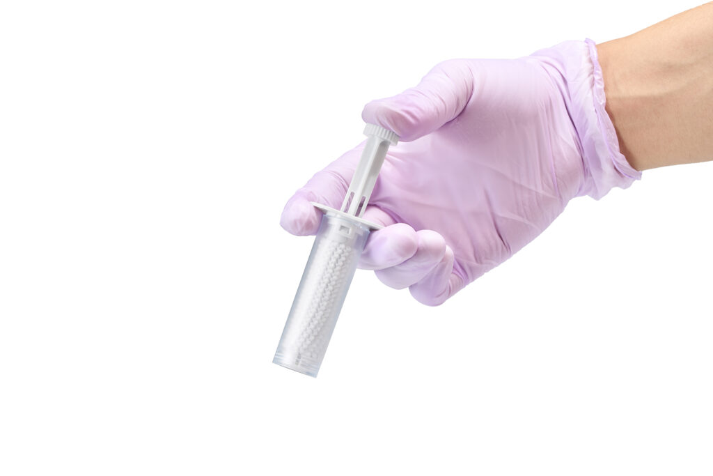
2. Transfer sample into the test cartridge and run it on the FLASH BaseTM
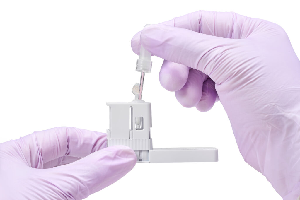
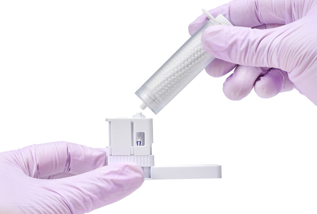
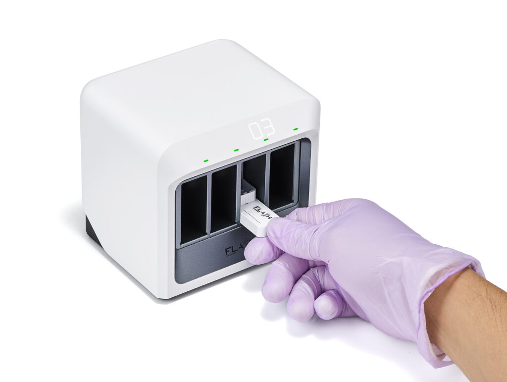
3. Activate the test cartridge and read the results
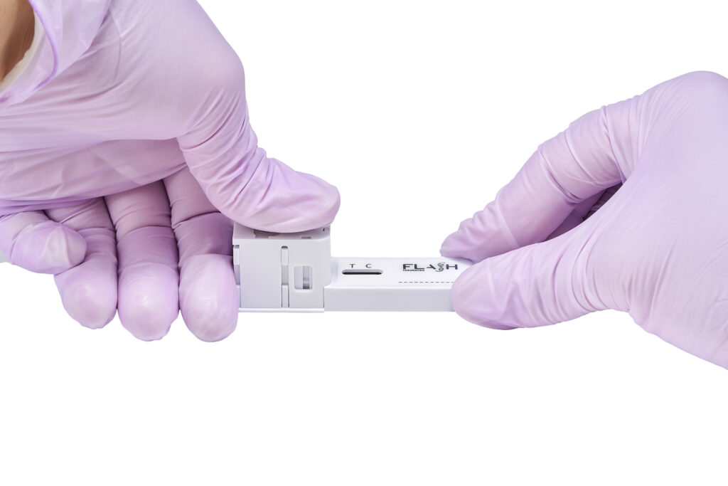
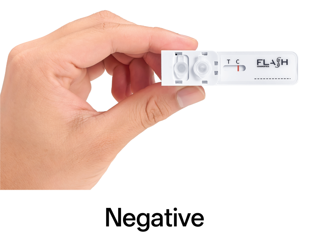
OR
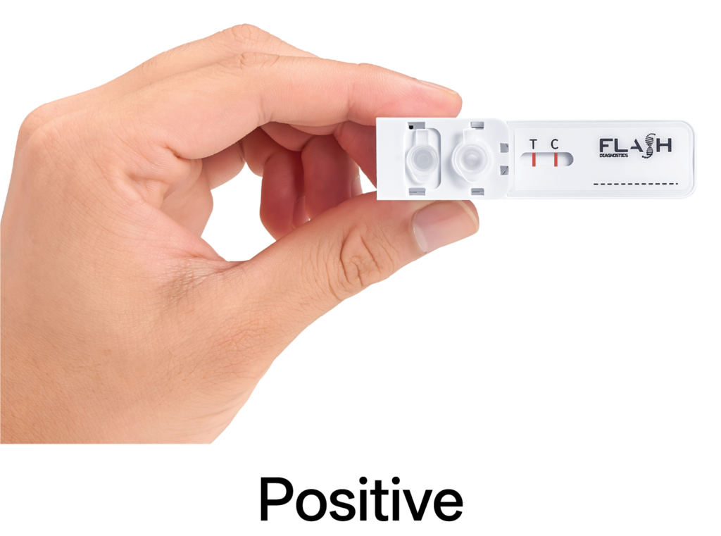
General information
Target: Ehrlichia canis
Species: Canine
Sample type: 50 μL EDTA blood
Result interpretation: Visual – lateral flow strip
Format: Single-use disposable test kit
Reagents: Lyophilized beads for isothermal amplification reaction
Hands-on sample preparation: 5 minutes + 5 minutes centrifugation
Reaction time: 30 minutes
Analytical limit of detection: 1,250 copies per reaction
Shelf life and storage: 12 months at room temperature